Ubiquitin B
Protein-coding gene in the species Homo sapiens
| UBB | |||||||||||||||||||||||||||||||||||||||||||||||||||
|---|---|---|---|---|---|---|---|---|---|---|---|---|---|---|---|---|---|---|---|---|---|---|---|---|---|---|---|---|---|---|---|---|---|---|---|---|---|---|---|---|---|---|---|---|---|---|---|---|---|---|---|
 | |||||||||||||||||||||||||||||||||||||||||||||||||||
| Identifiers | |||||||||||||||||||||||||||||||||||||||||||||||||||
| Aliases | UBB, HEL-S-50, Ubiquitin B | ||||||||||||||||||||||||||||||||||||||||||||||||||
| External IDs | OMIM: 191339; MGI: 98888; HomoloGene: 75104; GeneCards: UBB; OMA:UBB - orthologs | ||||||||||||||||||||||||||||||||||||||||||||||||||
| |||||||||||||||||||||||||||||||||||||||||||||||||||
| |||||||||||||||||||||||||||||||||||||||||||||||||||
| |||||||||||||||||||||||||||||||||||||||||||||||||||
| |||||||||||||||||||||||||||||||||||||||||||||||||||
| |||||||||||||||||||||||||||||||||||||||||||||||||||
| Wikidata | |||||||||||||||||||||||||||||||||||||||||||||||||||
| |||||||||||||||||||||||||||||||||||||||||||||||||||


Ubiquitin is a protein that in humans is encoded by the UBB gene.[5]
Function
Ubiquitin is one of the most conserved proteins known in eukaryotic organisms. Ubiquitin is required for ATP-dependent, non-lysosomal intracellular protein degradation of abnormal proteins and normal proteins with a rapid turnover. Ubiquitin is covalently bound to proteins to be degraded, and presumably labels these proteins for degradation. Ubiquitin also binds to histone H2A in actively transcribed regions but does not cause histone H2A degradation, suggesting that ubiquitin is also involved in regulation of gene expression. This gene consists of three direct repeats of the ubiquitin coding sequence with no spacer sequence. Consequently, the protein is expressed as a polyubiquitin precursor with a final amino acid after the last repeat. Aberrant form of this protein (UBB+1) has been noticed in patients with Alzheimer's disease, Down syndrome, other tauopathies (e.g. Pick's disease) and polyglutamine disease (e.g. Huntington's disease).[6][7]
References
- ^ a b c GRCh38: Ensembl release 89: ENSG00000170315 – Ensembl, May 2017
- ^ a b c GRCm38: Ensembl release 89: ENSMUSG00000019505 – Ensembl, May 2017
- ^ "Human PubMed Reference:". National Center for Biotechnology Information, U.S. National Library of Medicine.
- ^ "Mouse PubMed Reference:". National Center for Biotechnology Information, U.S. National Library of Medicine.
- ^ Webb GC, Baker RT, Fagan K, Board PG (Mar 1990). "Localization of the human UbB polyubiquitin gene to chromosome band 17p11.1-17p12". Am J Hum Genet. 46 (2): 308–15. PMC 1684968. PMID 2154095.
- ^ Fischer DF, De Vos RA, Van Dijk R, De Vrij FM, Proper EA, Sonnemans MA, Verhage MC, Sluijs JA, Hobo B, Zouambia M, Steur EN, Kamphorst W, Hol EM, Van Leeuwen FW (Nov 2003). "Disease-specific accumulation of mutant ubiquitin as a marker for proteasomal dysfunction in the brain". FASEB J. 17 (14): 2014–2024. doi:10.1096/fj.03-0205com. PMID 14597671. S2CID 10932825.
- ^ "Entrez Gene: UBB ubiquitin B".
Further reading
- Conaway RC, Brower CS, Conaway JW (2002). "Emerging roles of ubiquitin in transcription regulation". Science. 296 (5571): 1254–1258. Bibcode:2002Sci...296.1254C. doi:10.1126/science.1067466. PMID 12016299. S2CID 38822700.
- Murphey RK, Godenschwege TA (2002). "New roles for ubiquitin in the assembly and function of neuronal circuits". Neuron. 36 (1): 5–8. doi:10.1016/S0896-6273(02)00943-1. PMID 12367500. S2CID 15764136.
- Mazzé FM, Degrève L (2006). "The role of viral and cellular proteins in the budding of human immunodeficiency virus". Acta Virol. 50 (2): 75–85. PMID 16808324.
- Schlesinger DH, Goldstein G (1975). "Molecular conservation of 74 amino acid sequence of ubiquitin between cattle and man". Nature. 255 (5507): 423–4. Bibcode:1975Natur.255..423S. doi:10.1038/255423a0. PMID 1128706. S2CID 4159096.
- Adams SM, Sharp MG, Walker RA, et al. (1992). "Differential expression of translation-associated genes in benign and malignant human breast tumours". Br. J. Cancer. 65 (1): 65–71. doi:10.1038/bjc.1992.12. PMC 1977345. PMID 1370760.
- Pancré V, Pierce RJ, Fournier F, et al. (1991). "Effect of ubiquitin on platelet functions: possible identity with platelet activity suppressive lymphokine (PASL)". Eur. J. Immunol. 21 (11): 2735–41. doi:10.1002/eji.1830211113. PMID 1657614. S2CID 23901646.
- Baker RT, Board PG (1991). "The human ubiquitin-52 amino acid fusion protein gene shares several structural features with mammalian ribosomal protein genes". Nucleic Acids Res. 19 (5): 1035–1040. doi:10.1093/nar/19.5.1035. PMC 333777. PMID 1850507.
- Fornace AJ, Alamo I, Hollander MC, Lamoreaux E (1989). "Ubiquitin mRNA is a major stress-induced transcript in mammalian cells". Nucleic Acids Res. 17 (3): 1215–1230. doi:10.1093/nar/17.3.1215. PMC 331738. PMID 2537950.
- Lund PK, Moats-Staats BM, Simmons JG, et al. (1985). "Nucleotide sequence analysis of a cDNA encoding human ubiquitin reveals that ubiquitin is synthesized as a precursor". J. Biol. Chem. 260 (12): 7609–13. doi:10.1016/S0021-9258(17)39652-7. PMID 2581967.
- Einspanier R, Sharma HS, Scheit KH (1987). "Cloning and sequence analysis of a cDNA encoding poly-ubiquitin in human ovarian granulosa cells". Biochem. Biophys. Res. Commun. 147 (2): 581–587. doi:10.1016/0006-291X(87)90970-3. PMID 2820408.
- Wiborg O, Pedersen MS, Wind A, et al. (1985). "The human ubiquitin multigene family: some genes contain multiple directly repeated ubiquitin coding sequences". EMBO J. 4 (3): 755–9. doi:10.1002/j.1460-2075.1985.tb03693.x. PMC 554252. PMID 2988935.
- Baker RT, Board PG (1987). "The human ubiquitin gene family: structure of a gene and pseudogenes from the Ub B subfamily". Nucleic Acids Res. 15 (2): 443–463. doi:10.1093/nar/15.2.443. PMC 340445. PMID 3029682.
- Vijay-Kumar S, Bugg CE, Cook WJ (1987). "Structure of ubiquitin refined at 1.8 A resolution". J. Mol. Biol. 194 (3): 531–544. doi:10.1016/0022-2836(87)90679-6. PMID 3041007.
- Busch H (1984). "Ubiquitination of Proteins". Posttranslational Modifications Part A. Methods in Enzymology. Vol. 106. pp. 238–262. doi:10.1016/0076-6879(84)06025-0. ISBN 978-0-12-182006-0. PMID 6092831.
- Andersen MW, Ballal NR, Goldknopf IL, Busch H (1981). "Protein A24 lyase activity in nucleoli of thioacetamide-treated rat liver releases histone 2A and ubiquitin from conjugated protein A24". Biochemistry. 20 (5): 1100–1104. doi:10.1021/bi00508a009. PMID 6261785.
- Busch H, Goldknopf IL (1982). "Ubiquitin - protein conjugates". Mol. Cell. Biochem. 40 (3): 173–87. doi:10.1007/bf00224611. PMID 6275256. S2CID 21101636.
- Treier M, Staszewski LM, Bohmann D (1994). "Ubiquitin-dependent c-Jun degradation in vivo is mediated by the delta domain". Cell. 78 (5): 787–798. doi:10.1016/S0092-8674(94)90502-9. PMID 8087846. S2CID 33344164.
- Cook WJ, Jeffrey LC, Kasperek E, Pickart CM (1994). "Structure of tetraubiquitin shows how multiubiquitin chains can be formed". J. Mol. Biol. 236 (2): 601–609. doi:10.1006/jmbi.1994.1169. PMID 8107144.
- Ramage R, Green J, Muir TW, et al. (1994). "Synthetic, structural and biological studies of the ubiquitin system: the total chemical synthesis of ubiquitin". Biochem. J. 299 (1): 151–8. doi:10.1042/bj2990151. PMC 1138034. PMID 8166633.
- v
- t
- e
PDB gallery
-
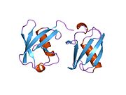
 1aar: STRUCTURE OF A DIUBIQUITIN CONJUGATE AND A MODEL FOR INTERACTION WITH UBIQUITIN CONJUGATING ENZYME (E2)
 -
 1cmx: STRUCTURAL BASIS FOR THE SPECIFICITY OF UBIQUITIN C-TERMINAL HYDROLASES
 -
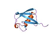
 1d3z: UBIQUITIN NMR STRUCTURE
 -
 1f9j: STRUCTURE OF A NEW CRYSTAL FORM OF TETRAUBIQUITIN
 -
 1fxt: STRUCTURE OF A CONJUGATING ENZYME-UBIQUITIN THIOLESTER COMPLEX
 -
 1g6j: STRUCTURE OF RECOMBINANT HUMAN UBIQUITIN IN AOT REVERSE MICELLES
 -
 1gjz: SOLUTION STRUCTURE OF A DIMERIC N-TERMINAL FRAGMENT OF HUMAN UBIQUITIN
 -
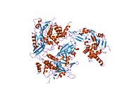 1nbf: Crystal structure of a UBP-family deubiquitinating enzyme in isolation and in complex with ubiquitin aldehyde
 -
 1ogw: SYNTHETIC UBIQUITIN WITH FLUORO-LEU AT 50 AND 67
 -
 1otr: Solution Structure of a CUE-Ubiquitin Complex
 -
 1p3q: Mechanism of Ubiquitin Recognition by the CUE Domain of VPS9
 -
 1q0w: Solution structure of Vps27 amino-terminal UIM-ubiquitin complex
 -
 1q5w: Ubiquitin Recognition by Npl4 Zinc-Fingers
 -
 1s1q: TSG101(UEV) domain in complex with Ubiquitin
 -
 1sif: Crystal structure of a multiple hydrophobic core mutant of ubiquitin
 -
 1tbe: STRUCTURE OF TETRAUBIQUITIN SHOWS HOW MULTIUBIQUITIN CHAINS CAN BE FORMED
 -
 1ubi: SYNTHETIC STRUCTURAL AND BIOLOGICAL STUDIES OF THE UBIQUITIN SYSTEM. PART 1
 -
 1ubq: STRUCTURE OF UBIQUITIN REFINED AT 1.8 ANGSTROMS RESOLUTION
 -
 1ud7: SOLUTION STRUCTURE OF THE DESIGNED HYDROPHOBIC CORE MUTANT OF UBIQUITIN, 1D7
 -
 1uzx: A COMPLEX OF THE VPS23 UEV WITH UBIQUITIN
 -
 1v80: Solution structures of ubiquitin at 30 bar and 3 kbar
 -
 1v81: Solution structures of ubiquitin at 30 bar and 3 kbar
 -
 1wr1: The complex structure of Dsk2p UBA with ubiquitin
 -
 1wr6: Crystal structure of GGA3 GAT domain in complex with ubiquitin
 -
 1wrd: Crystal structure of Tom1 GAT domain in complex with ubiquitin
 -
 1xd3: Crystal structure of UCHL3-UbVME complex
 -
 1xqq: Simultaneous determination of protein structure and dynamics
 -
 1yd8: COMPLEX OF HUMAN GGA3 GAT DOMAIN AND UBIQUITIN
 -
 1yiw: X-ray Crystal Structure of a Chemically Synthesized Ubiquitin
 -
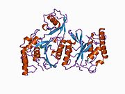
![1yj1: X-ray Crystal Structure of a Chemically Synthesized [D-Gln35]Ubiquitin](//upload.wikimedia.org/wikipedia/commons/thumb/f/f5/PDB_1yj1_EBI.jpg/180px-PDB_1yj1_EBI.jpg) 1yj1: X-ray Crystal Structure of a Chemically Synthesized [D-Gln35]Ubiquitin
1yj1: X-ray Crystal Structure of a Chemically Synthesized [D-Gln35]Ubiquitin -
 1yx5: Solution Structure of S5a UIM-1/Ubiquitin Complex
 -
 1yx6: Solution Structure of S5a UIM-2/Ubiquitin Complex
 -
 1zgu: Solution structure of the human Mms2-Ubiquitin complex
 -
 2ayo: Structure of USP14 bound to ubquitin aldehyde
 -
 2bgf: NMR STRUCTURE OF LYS48-LINKED DI-UBIQUITIN USING CHEMICAL SHIFT PERTURBATION DATA TOGETHER WITH RDCS AND 15N-RELAXATION DATA
 -
 2c7m: HUMAN RABEX-5 RESIDUES 1-74 IN COMPLEX WITH UBIQUITIN
 -
 2c7n: HUMAN RABEX-5 RESIDUES 1-74 IN COMPLEX WITH UBIQUITIN
 -
 2d3g: Double sided ubiquitin binding of Hrs-UIM
 -
 2den: Solution Structure of the Ubiquitin-Associated Domain of Human BMSC-UbP and its Complex with Ubiquitin
 -
 2dx5: The complex structure between the mouse EAP45-GLUE domain and ubiquitin
 -
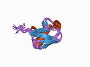
![2fcm: X-ray Crystal Structure of a Chemically Synthesized [D-Gln35]Ubiquitin with a Cubic Space Group](//upload.wikimedia.org/wikipedia/commons/thumb/1/1a/PDB_2fcm_EBI.jpg/180px-PDB_2fcm_EBI.jpg) 2fcm: X-ray Crystal Structure of a Chemically Synthesized [D-Gln35]Ubiquitin with a Cubic Space Group
2fcm: X-ray Crystal Structure of a Chemically Synthesized [D-Gln35]Ubiquitin with a Cubic Space Group -
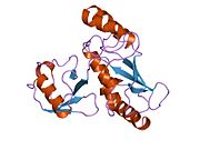
![2fcn: X-ray Crystal Structure of a Chemically Synthesized [D-Val35]Ubiquitin with a Cubic Space Group](//upload.wikimedia.org/wikipedia/commons/thumb/d/d8/PDB_2fcn_EBI.jpg/180px-PDB_2fcn_EBI.jpg) 2fcn: X-ray Crystal Structure of a Chemically Synthesized [D-Val35]Ubiquitin with a Cubic Space Group
2fcn: X-ray Crystal Structure of a Chemically Synthesized [D-Val35]Ubiquitin with a Cubic Space Group -
 2fcq: X-ray Crystal Structure of a Chemically Synthesized Ubiquitin with a Cubic Space Group
 -
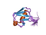
![2fcs: X-ray Crystal Structure of a Chemically Synthesized [L-Gln35]Ubiquitin with a Cubic Space Group](//upload.wikimedia.org/wikipedia/commons/thumb/e/e6/PDB_2fcs_EBI.jpg/180px-PDB_2fcs_EBI.jpg) 2fcs: X-ray Crystal Structure of a Chemically Synthesized [L-Gln35]Ubiquitin with a Cubic Space Group
2fcs: X-ray Crystal Structure of a Chemically Synthesized [L-Gln35]Ubiquitin with a Cubic Space Group -
 2fid: Crystal Structure of a Bovine Rabex-5 fragment complexed with ubiquitin
 -
 2fif: Crystal Structure of a Bovine Rabex-5 fragment complexed with ubiquitin
 -
 2fuh: Solution Structure of the UbcH5c/Ub Non-covalent Complex
 -
 2g3q: Solution Structure of Ede1 UBA-ubiquitin complex
 -
 2g45: Co-crystal structure of znf ubp domain from the deubiquitinating enzyme isopeptidase T (isot) in complex with ubiquitin
 -
 2gbj: Crystal Structure of the 9-10 8 Glycine Insertion Mutant of Ubiquitin.
 -
 2gbk: Crystal Structure of the 9-10 MoaD Insertion Mutant of Ubiquitin
 -
 2gbm: Crystal Structure of the 35-36 8 Glycine Insertion Mutant of Ubiquitin
 -
 2gbn: Crystal Structure of the 35-36 8 Glycine Insertion Mutant of Ubiquitin
 -
 2gbr: Crystal Structure of the 35-36 MoaD Insertion Mutant of Ubiquitin
 -
 2gmi: Mms2/Ubc13~Ubiquitin
 -
 2hd5: USP2 in complex with ubiquitin
 -
 2hth: Structural basis for ubiquitin recognition by the human EAP45/ESCRT-II GLUE domain
 -
 2ibi: Covalent Ubiquitin-USP2 Complex
 -
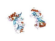
 2j7q: CRYSTAL STRUCTURE OF THE UBIQUITIN-SPECIFIC PROTEASE ENCODED BY MURINE CYTOMEGALOVIRUS TEGUMENT PROTEIN M48 IN COMPLEX WITH A UBQUITIN-BASED SUICIDE SUBSTRATE
 -
 2nr2: The MUMO (minimal under-restraining minimal over-restraining) method for the determination of native states ensembles of proteins
 -
 2o6v: Crystal structure and solution NMR studies of Lys48-linked tetraubiquitin at neutral pH
 -
 2oob: crystal structure of the UBA domain from Cbl-b ubiquitin ligase in complex with ubiquitin


![1yj1: X-ray Crystal Structure of a Chemically Synthesized [D-Gln35]Ubiquitin](http://upload.wikimedia.org/wikipedia/commons/thumb/f/f5/PDB_1yj1_EBI.jpg/180px-PDB_1yj1_EBI.jpg)


![2fcm: X-ray Crystal Structure of a Chemically Synthesized [D-Gln35]Ubiquitin with a Cubic Space Group](http://upload.wikimedia.org/wikipedia/commons/thumb/1/1a/PDB_2fcm_EBI.jpg/180px-PDB_2fcm_EBI.jpg)
![2fcn: X-ray Crystal Structure of a Chemically Synthesized [D-Val35]Ubiquitin with a Cubic Space Group](http://upload.wikimedia.org/wikipedia/commons/thumb/d/d8/PDB_2fcn_EBI.jpg/180px-PDB_2fcn_EBI.jpg)

![2fcs: X-ray Crystal Structure of a Chemically Synthesized [L-Gln35]Ubiquitin with a Cubic Space Group](http://upload.wikimedia.org/wikipedia/commons/thumb/e/e6/PDB_2fcs_EBI.jpg/180px-PDB_2fcs_EBI.jpg)


 | This article on a gene on human chromosome 17 is a stub. You can help Wikipedia by expanding it. |
- v
- t
- e


